Homeowners at Tengah EC may be asking what are green features and why they should consider installing them in their property. A simple understanding of what a feature entails will help to answer this question. In North America, most homeowners understand that green features are those items that can be recycled or used, and some even choose to sell the product of their green features. These features can be anything from trees to grass clippings and more. However, not everyone knows that some of the green features available on the market today can actually replace more traditional materials such as stone, brick, concrete, and asphalt.
An example of a green landscape feature is grass clippings at Tengah EC. The process of collecting these clippings, as well as making small plots of land around each clippings will provide plant life for the plants. The lawn will look its best, and the plants will also grow faster than they would without the grass clippings. This landscape feature is similar to what you might see on a compost heap, but instead of material coming out of the pile, plant life will be growing within the pile. This allows the homeowner to use less soil, and even more grass clippings if desired. This is a very simple process and all the landscape architect needs to do is put in a sprinkler system to get the clippings to the ground.
Another landscape feature that is considered green is the use of wood mulch. Mulch is made from natural materials that are not harmful to the environment. This type of feature can be installed anywhere on your property, but some recommend placing it under the trellis poles, trees, gazebos, and other structures that can be found on your property. This feature will help to reduce weeds and protect the soil, which in turn improves water quality. This reduces the amount of water used by the landscaping company when the work is finished.
Using green features doesn’t mean that the landscape architect has to design the feature all by himself at Tengah EC Choa Chu Kang. Many landscape companies offer feature kits that can be used at any construction site. These feature kits include everything, the landscape architect needs to complete the project. Some feature kits even include the sand and stone for the feature. These kits make it easy to select and install the different green features on the landscape.
Landscape companies often offer these features because they are inexpensive and require less effort than other options. Some of the green features use recycled materials to save money, but many use natural materials. Recycled materials include cork tiles, crushed granite, and even coconut timbers, which were originally used in Bali.
When choosing a green feature at Tengah EC, it’s important to consider the location of the feature, how it will be installed, and how it will look once complete. All of these factors play a role in the price of the feature. For example, if the feature is to be located on top of a concrete slab, the price may be higher because of the cost of tearing down the concrete. The placement of the feature is also important. If a feature is too close to existing trees or other features, the cost of landscaping the area may be greater than simply using a recycled material.
When using recycled materials for a landscape feature, using the green features can help the architect save a considerable amount of money. Some of these features can be constructed from recycled materials, such as cork tile. Some designers choose to purchase cork tiles directly from demolition companies. Another way an architect can save money on green features is to have the feature built with reused materials. This often happens when an architect designs an addition to an existing house rather than designing the whole new house from scratch.
An added benefit of incorporating green features into a landscape architect’s design is that it makes the landscape more beautiful and the home more comfortable. If this feature is installed correctly, the architect can use the natural materials available to construct the feature. The combination of the use of natural materials and creative thinking helps to make the finished product a more pleasing experience.
Yishun EC, or Yishun City, is one of the most promising areas in the North of Singapore. Located right in the center of Yishun, it is conveniently close to various facilities at the heart of Yishun, and also to most attractions in Yishun itself. The development is just a short walk away from Yishun MRT station and is situated on Yishun Avenue, 9. It is one of the most upcoming developments in Phuket and boasts numerous benefits.
The Yishun Integrated Transport Hub in Northpoint City provides a good service and allows tourists to travel around the region using the world’s most advanced public transportation system, the MRT. But the best part about this shopping and eating center is that it is within walking distance to the Yishun EC and has an integrated mall. The mall includes a food court, a wide variety of restaurants, and a variety of retail shops.
This mall is conveniently located near the Yishun MRT Station and the major bus stations. There are several hotels located nearby, including the Yishun Lanes and the Yishun International Airport. The Yishun International Airport offers flights to Thailand, as well as other Asian countries. This means that visitors can easily make it into the city and head straight to their hotel, without having to take a cab or spend much time traveling through the city. If they don’t want to leave the airport, they can simply take the bus and drive themselves to their hotel, which is conveniently located near all the amenities.
One of the facilities offered at this shopping centre is the Yishun EC Mall. This mall features around 300 shops, restaurants and food courts. It is also near the Yishun MRT station and the northpoint city center.
The Yishun EC Shopping Centre is located just beside the Singapore River. This means that visitors can walk to the EC Shopping Centre from anywhere in the north. There is no need to taxi or ride the metro to get here. The sky bridge over the Singapore River gives easy access to this shopping centre. The shops here sell everything from local products to toys, clothes, books, jewelries and electronic goods.
Another popular structure located near Yishun EC are the Yishun Residences. These apartments are one of the best resale properties in the country and offer luxurious living standards at affordable prices. The majority of these residences are sold by a group of builders who have built these private residential properties to be sold as a fully furnished, fully managed and operated private town home community.
Yishun Residences is also located close to some of the best shopping malls in the country. These shopping malls include the Yishun International Complex, the Singapore Plaza, the Big Bazaar, and the Yishun International Mall. These shopping malls are known for their modern and sophisticated design. The majority of these malls have their own restaurants. Visitors can take a lunch inside the shopping mall and then drive out towards the popular night spots.
Some of the best features offered by Yishun Properties are its location, its design, its layout, and the quality of its construction materials. Yishun properties are in high demand and have rapidly risen in value. Its proximity to prominent Singapore landmarks and the shopping malls it is situated near make it one of the most sought after Singapore residential properties today. This prime piece of real estate is worth every penny, whatever its price tag. Given the current condition of the economy, a buyer can practically afford a Yishun EC, even with today’s depressed prices.
Located only a few minutes walk from Yishun Industrial Estate, the residents of Yishun EC can enjoy all the benefits that come with a modern lifestyle. This premier Singapore residential property is conveniently located, making it easy for residents to commute to work in the city center. Those looking for affordable living can opt for this luxurious and spacious Singapore residential estate.
Located in the heart of the north east suburbs, Yishun EC gives residents all the convenience of an urban lifestyle without the traffic hassle. In just minutes, one can reach the Yishun MRT Station. The MRT is the preferred means of traveling between the east coast and the business centers of the business capital of Singapore. It is also just minutes away from the popular Singapore beaches such as the Orchard Road, the Singapore Botanic Garden, the Victoria Peak, and the Yishun International Airport. When a resident is so close to all the major attractions of the city, shopping should not be a problem. Shopping in Singapore is a very popular past time and some of the popular shopping districts of the country are Central Business District, Orchard Road and the Central Business Districts of the country.
For those who prefer to live in a condominium, Yishun EC offers an exclusive lifestyle for its residents. The group developed this residential estate to cater to the needs of its residents. Unlike other luxury residential properties, Yishun EC offers its residents a high standard of security and privacy. These features make it a perfect place for newlywed couples to start their married life in Singapore.
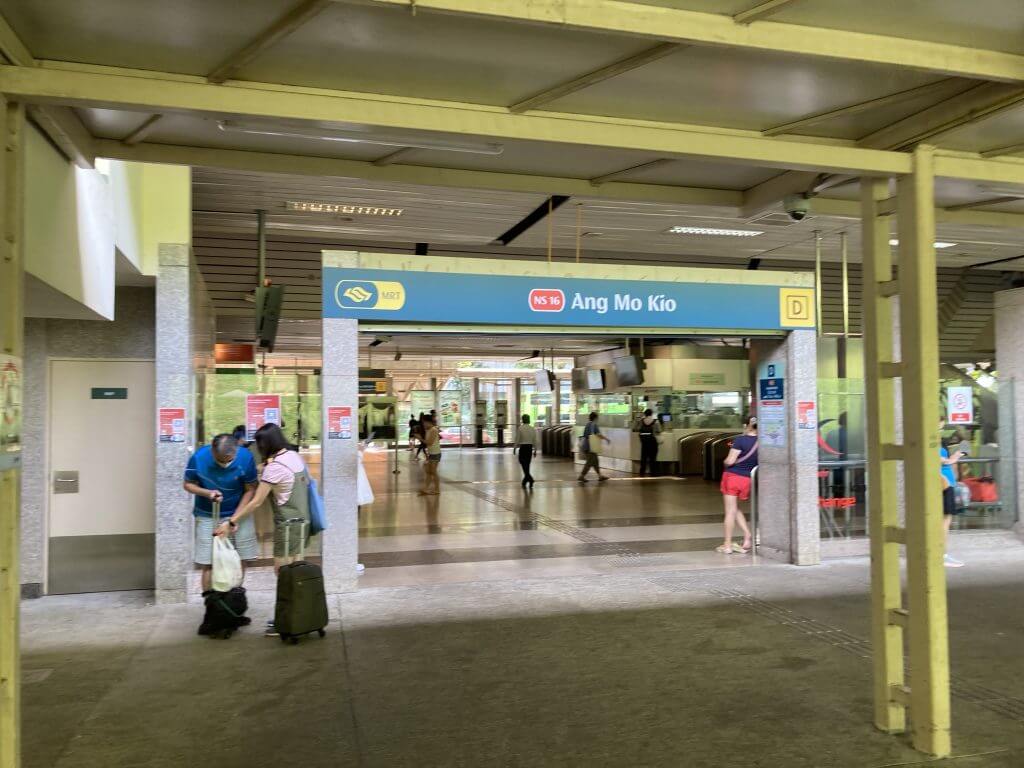
The five star luxury villa in Singapore, Belgravia Ace Ang Mo Kio promises spectacular views over the city skyline and is located in the heart of the business district of central Singapore. Belgravia Ace villa is nestled at the end of a narrow sloping driveway and is surrounded by lush gardens bursting with tropical flowers. Belgravia Ace is designed with an ultra-modern aesthetic appeal, incorporating cutting-edge furniture, sleek glass appliances and exposed brick walls. Belgravia Ace property has been landscaped with scenic gardens and features two swimming pools with water slides, as well as a spacious sun deck. Belgravia Ace property is also provided with high speed internet access and an onsite fitness centre.
Belgravia Ace Ang Mo Kio Development Village is a unique mixed use development that utilises both the city’s existing public transport infrastructure, as well as its own transport facilities. Belgravia Ace village offers residents easy access to the underground train station, the airport, and Central Business District (CBD). The buildings at the site are designed to cater for an international community who may be interested in investing in a home here. Most of Belgravia Ace villas in the development are designed with a dual purpose. They offer residents convenient access to their own private homes, as well as the amenities they require to live a comfortable lifestyle. The buildings at Ang Mo Kio are designed with two levels, each offering residents with their own private terrace.
Belgravia Ace Semi Detached and Terrace Houses Available for Launch soon by Tong Eng Group
Semi Detached and Terrace Houses landscape design of Belgravia Ace Ang Mo Kio Village, is constructed around an artificial lagoon, providing a relaxing and tranquil setting for residents. A landscaped tropical garden completes the landscape design and includes a pond and a water play area for the kids. Several small roads, as well as pathways, allow residents to move between their private residential homes and the landscaped garden area. This gives residents an opportunity to mingle with other private residential owners and families.
Belgravia Ace Ang Mo Kio Avenue 5
The Belgravia Ace in Ang Mo Kio are all very luxurious Semi Detached and Terrace Houses . Most of Belgravia Ace offer round the clock room service, as well as satellite television. Belgravia Ace rooms feature fully furnished kitchens, with modern kitchen appliances, including blenders, dishwashers, and ovens. Several of the Belgravia Ace feature private decks that are accessible via their own unique access points, while some of the Ang Mo Kio Belgravia Ace also have shared decks. Belgravia Ace private residential areas of Ang Mo Kio are designed around a central garden. The landscaped garden area includes a pond, a water play area for children, and several pavilions. There is also a public landscaped garden, which offers an assortment of tropical plants and flowering trees. Many of the gardens feature bird houses, which are supported by steel posts. Belgravia Ace gardens are designed to be relaxing, as well as entertaining. The private residential areas at Ang Mo Kio are secluded, private, and easy to access from the airport.
Belgravia Ace Ang Mo Kio, one can find a private golf course, secluded leisurely beach access, and a public tennis court. There is also a secondary school, St Joseph’s School, which offers a curriculum that is aligned with the Australian Standard. The secondary school features an Islamic studies curriculum, and students are required to participate in extra-curricular activities, such as playing basketball. The secondary school is only for boys. Belgravia Ace gardens at Ang Mo Kio are designed to be an extension of the villas, and are surrounded by beautiful scenery. There are wide walkways throughout the gardens, which offer visitors the chance to walk among the different types of flora and fauna. At one end of the gardens is a small observation deck that overlooks the water. Further along are the Serangoon and Mo Jui Water Bodies, which are considered to be Australia’s largest and most impressive water bodies.
In addition to the gardens, Belgravia Ace Ang Mo Kio offers many luxurious features. The five restaurants and five-star hotels are located at the center of the garden area, surrounded by lush landscaping. The five-star hotel features a swimming pool, and all of the restaurants feature delicious local and international cuisine. The second largest city in the state of Singapore, Belgravia Ace is also known as “The Jewel of Singapore”. With its rich heritage, the city has a diverse population spread in all levels and demographics. A UNESCO World Heritage site, the city is home to a large number of national and international attractions. One of the best things to do in Belgravia Ace is to stay in one of the many villas here.
Belgravia Ace Semi Detached and Terrace Houses Available for Launch soon by Tong Eng Group
A visit to this Semi Detached Houses is always worthwhile because of its natural beauty. As you travel towards the city, you will be greeted by the enchanting scenery of Singapore’s Mount Cook – a picturesque promenade that peaks at about 300 meters. In this secluded area, you can relax, take in the breathtaking view of the city, and indulge in a cup of delicious coffee. Belgravia Ace view from here is quite extraordinary because of its unusual shape. The topography of this landscape design features steep cliffs and grassy slopes covered with thick vegetation. Belgravia Ace water features here are very unique and offer several interesting attractions. On your way to Ang Mo Kio, look out your left and right depending on what path you wish to take. Belgravia Ace begins from the middle of the road and curves around the bend to a path just beside a tree-studded hillside. From there, you will see a picturesque landscape that offers a panoramic view of the reservoir and the surrounding gardens. At this point, you will notice the steepest part of the Ang Mo Kio trek which is lined with trees – which provide a sanctuary for birds and wildlife. At the end of the trek, you will discover a plaque commemorating Ang Mo Kio and the first discovery of the Western Ghats in Sri Lanka.
Once you reach the end of the Ang Mo Kio trek, turn left and proceed to visit Ang Mo Kio’s Semi Detached and Terrace Houses . In these waterfronts, you will find the Ang Mo Kio reservoir and the observation deck. In addition to water and a place to sit, you can have your photograph taken on the observation deck at any time – it is all included in the Ang Mo Kio flatland suites or flats. You will find another popular attraction in Belgravia Ace Ang Mo Kio. Known as seletar aerospace park, this is where a commercial airline operates their aircraft. To get to this tourist attraction, you must use Ang Mo Kio’s primary airport, Batangala. Seletar is also served by a number of private residential communities. Many of these residents own holiday home accommodation on the site of Ang Mo Kio.
For those who are interested in visiting the Serangoon National Park, you must visit Belgravia Ace Ang Mo Kio Secondary School. This school offers a wide selection of learning experiences to its students. The school is situated next to the selector jetty. The Serangoon National Park is known for its tropical forests, rich flora and fauna, wildlife and other interesting natural attractions. A visit here will not be complete without a walk around the forest, just to get a feel of what the place has to offer. A visit to the Serangoon National Park is not complete without a visit to the Ang Mo Kio Golf Course. Here you can practice golf and spend your days exploring the rich natural vegetation and scenery of this scenic area. There is a small village nearby the golf course, which offers a few houses and an open air restaurant.
Belgravia Ace Semi Detached and Terrace Houses Available for Launch soon by Tong Eng Group
If you wish to have some fun during your stay in Belgravia Ace and want to explore more of the deluxe accommodation facilities available in the area, then you can opt for a holiday villa in Belgravia Semi Detached. You may stay in Belgravia Ace available at Tanginoa. You will find that the surroundings are quite picturesque, with a pleasant setting. Most of the holiday villas in Belgravia are serviced with two swimming pools and the facilities to entertain family and friends. In addition to the holiday homes in Belgravia Ace, there are also a variety of private residential properties in Tanginoa. Belgravia Ace Ang Mo Kio is located in close proximity to Kippong and the entertainment district of Singapore. Tourists in search for authentic Asian foods will find that this location has a wide range of restaurants to offer. The variety of cuisine is very interesting, ranging from spicy Indian, Portuguese, Peranakan, Chinese, Lebanese and other local specialties. The most popular dishes are Halal meat, Garlic Pepper Steak, Massaman curry, Mian ka Farang, Chicken Rice, Laksa. These restaurants also serve other delicacies such as desserts such as banana shake, malai melon and many more.
The authentic Thai food is served at this restaurant. They offer food from every part of Asia, starting from India and ending with China. They also have pizza and Chinese barbecues. This restaurant is located just next to the Night Markets, where you can find a wide variety of affordable Chinese and native cuisine. Nang Oe restaurant is located in the northern part of Kippong. This is one of the original Japanese restaurants that serve authentic Japanese food. They offer dishes such as Japanese sushi, beef sashimi, chicken dishes and seafood such as crab and octopus. They also offer a high quality of shopping, ranging from apparel and jewelry to electronics and accessories.
Semi Detched and Terrace Houses Landed Property
In addition to the many Japanese restaurants, Belgravia Ace Semi Detached and Terrace Houses location offers many other specialty restaurants such as Pacho Fresco’s. This restaurant offers fresh seafood daily. They also serve a wide variety of desserts and snacks. Some of their entrees include fried squid, crab and lobster bisque, crab cakes, chicken pho and egg drop soup. There are also a variety of Japanese beers and wines on offer. If you are looking for authentic Chinese food in Kippong, you will not be disappointed with Chow Moi Restaurant. Belgravia Ace restaurant is located in a quiet corner of Ang Mo Kio. This is a traditional Chinese restaurant that serves delicious dim sum and delicious vegetarian dishes. Some of their entrees are steamed tenderloin, roast lamb, and fried dumplings. You can find some great seafood in the evening at this restaurant. You can even find a variety of Chinese desserts here.
Belgravia Ace Semi Detached and Terrace Houses Available for Launch soon by Tong Eng Group
Besides these two famous restaurants, there are many more restaurants located in Ang Mo Kio. You can choose from local eateries that offer affordable meals. Or, you can opt for high quality international food. These restaurants are located all across the city and offer many different cuisines. If you love sushi, you will not be disappointed by the wide selection of Asian eateries in Ang Mo Kio. In fact, you can even sit down and order just about any type of sushi during your stay in Kippong. You can find many Japanese restaurants located here. Some of these restaurants also offer Chinese food. The best way to see all of the different types of restaurants in Ang Mo Kio is to check them out when you come to town.
Belgravia Ace Ang Mo Kio is a great place to eat at any time of the day. There are always people walking around selling good food. If you want to find a great place to eat, you will definitely want to check out Ang Mo Kio. A lot of the restaurants in Belgravia Ace Ang Mo Kio are family owned. This is because the area was originally a rice field. There are many restaurants located here that cater to a variety of different tastes. The younger people in the area tend to be into burger and pizza. However, there are also many Asian restaurants that offer affordable meals. Whatever you like, you should definitely try some of the restaurants here.
There are many hotels in Ang Mo Kio as well. These hotels are a little more expensive than the typical hotels. Semi Detached and Terrace Houses rooms are usually clean, the staff is friendly, and the prices are fair. In addition, many of these hotels are located right on the water. This will allow you to take a boat cruise through the waters of Ang Mo Kio. If you are looking for a good restaurant, you should visit Sang Takla Food Court near Belgravia Ace. This restaurant is located inside the Ang Mo Kio International Airport. It is one of the best restaurants in the area and it offers a wide variety of food. The food is very reasonably priced and you should definitely try it out when in the area. Tong Eng Brothers Singapore is a leading real estate developer based in the country’s most cosmopolitan island, with locations in both the East and West Coast. They have several real estate developments in the high end Central Business Districts of the city such as Belgravia Ace. The company has built some very attractive commercial properties in the area including the prestigious shopping complex in the now defunct Remery Plaza mall.
Their other developments include luxury high-rise apartment blocks in the swanky Singapore Chinatown, Singapore Off Market and Singapore Botanical like Belgravia Ace. The company also has a number of retail outlets in the popular MRT station, the well-known Yean Yang Square and a string of posh malls including the Malls on Orchard Road and the Parco Marina. Their real estate developer’s portfolio is full of prime assets in prime locations across Asia and they regularly rank high in property surveys. Tong Eng Brothers has many partners in the country that span national and international expertise. Belgravia Ace stable infrastructure, knowledge and reputation for building great projects and partnering up with leading retailers and suppliers, creates a very attractive environment for buyers and sellers.
Belgravia Ace Semi Detached and Terrace Houses Available for Launch soon by Tong Eng Group
However, being the largest real estate developer in the country makes it difficult for them to compete with the other real estate developers. Competition for funding is stiff and even their partners are competing against each other for larger profits. Ongoing expansion into high-end facilities is taking its toll on their balance sheet and slowing down their growth. In response Tong Eng Brothers Singapore have launched a series of initiatives aimed at improving their standing in the market. These initiatives are focused on reducing cost and improving the overall performance of their real estate development portfolio.
Most of the buzz surrounding Tong Eng Brothers is centered around their partnership with the world’s largest furniture manufacturer, Beni Ourain for Belgravia Ace. This partnership has created a new segment within the company known as the ‘Beni Ourain Singapore Brand’. This brand is exclusively available through Tong Eng Brothers’ real estate developments in the country. The focus of this brand is to provide premium furniture at affordable prices to local buyers. Since launching in 2021, Beni Ourain has grown into one of the largest and most successful companies in the world.
Tong Eng Brothers Singapore also holds extensive partnerships with various other private companies. These partnerships focus on creating a diversified portfolio and strengthening the company’s position in the Singapore market. These partnerships have helped them expand into some of the more affluent locales of the country such as Jemma, Serang, and Bedok. They have also established joint ventures with several other well-known international real estate developers such as Zaha Hadid Architects and Urbancore.
At the ground level, Tong Eng Brothers Singapore have been working on solidifying their core competency in the real estate industry by continuously improving their products and services. They offer a wide variety of residential and commercial spaces in almost all price ranges. They have also made sure that they are constantly seeking out new opportunities and trends in the industry for Belgravia Ace. They have invested a lot of time and money in research and development. Their main areas of focus are residential spaces, commercial spaces, and infrastructure development projects. Although their residential product line has been developed primarily for local buyers, they also carry a selection of luxury properties that appeal to a global buyer base.
Located near the central business district of Singapore, Beni Ourain is one of the Tong Eng Brothers’ most important projects thus far. The development is composed of five townships and is surrounded by an expanse of landscaping and trees that create a tranquil atmosphere. Although the project is still in the early stages of development, already the scale of the real estate project is unprecedented in the small ring of suburbs around the city.
Tong Eng Brothers Singapore real estate developer is also responsible for developing another important property located within the ring, known as Belgravia Ace. Unlike the Beni Ourain project, this apartment complex is located adjacent to a major transportation hub in the area in Ang Mo Kio, meaning that frequent travelers into the country will be able to get to their destinations without having to take long detours through other less populated regions. Belgravia Ace provides a convenience for travelers who do not want to have to deal with navigating congested traffic. Another benefit of the New Britain Residences is that it is conveniently located near the Tong Eng Brothers Singapore offices. It is expected to be ready for occupancy in the first quarter of 2021.
Belgravia Ace is right in the middle of both activity and heaven: just 3 minutes away to the Central Expressway (CTE), easily accessible by means of bus quits and also major roads, and is additionally in the middle of an isolated area with a vast environment-friendly open space.There is a bus stop outside which serves the path Bishan, Ang Mo Kio, Sengkang, Hougang, and Upper Serangoon. Ang Mo Kio MRT Station on the North-South Line is likewise a couple of minute drive away, while the city correct is just a 12 to 15-minute drive.
There is additionally a wealth of nearby malls and dining establishments. Buying choices include Hougang 1, Greenwich V, Ang Mo Kio Center, MyVillage at Serangoon Garden, and also Seletar Shopping mall, which is connected to the Fernvale LRT station. A few of the eating choices nearby are Dragon Bowl, Pietro Ristorante Italiano, First Culinary Dining Establishment, and Seletar Hill Dining Establishment. The estate is additionally near Sheng Siong Hypermarket at Serangoon North, Titan, and NTUC Fairprice. Parents require not fret about their youngsters’s education, as this strata landed growth is near Hougang Primary School, Rosyth Institution, as well as Fernvale Primary School.
Belgravia Ace Landed Property Near to Central Expressway CTE at Ang Mo Kio Near to Greenwich V and Hougang 1
Other reputable educational institutions in the location are Chatsworth Preschool, Lycee Francais de Singapour (International French College), Dimensions International University, Anderson Serangoon Junior College, James Chef College, Anderson High School, Pei Hwa High School, Serangoon Yard Senior High School, Da Qiao Primary School, Nanyang Polytechnic, and also ITE University Central. It is also close to the 320-hectare Seletar Aerospace Park, an industrial park which was constructed to support the aerospace market. The park also has a number of eating alternatives, such as The Summerhouse and Wheelers Estate. The park additionally often hosts marketplace and also cultural occasions, as it houses a number of conserved colonial homes that were transformed right into a way of living haven. Finally, this freehold advancement is established by Fairview Dope Pte Ltd, a subsidiary of the recognized and also reliable Tong Eng Group.
Belgravia Ace is galvanized by the fashionable and also very preferred Belgravia area in Central London Belgravia Villas, which is equally set amongst a peaceful landed development steeped in culture as well as background. Located among a big site space of over 2 hectares among immense landscaping public open area, Belgravia Ace includes a special selection of 3 strata balcony houses and 104 semi-detached residences. Belgravia Ace is finely fitted out with noteworthy and polished devices and also installations from Italian manufacturer, De Dietrich and also Smeg whereas every of the five washrooms associate with hygienic wares from Villeroy and also Boch and premium faucets and also shower systems from Grohe in addition to your personal residence elevator, accessibility to your 5 storey of your home, consisting of 5 rooms with 4 en-suite washrooms, a washroom, a basement with 2 parking area, a roofing system terrace including wet and dry kitchen areas with a counter top.
Belgravia Ace is a treasure to discover, located on a mild hillside incline facing a classy 100m barrier with children’s playground next to it. Belgravia Villas is sitting on a big land dimension of more than 400,000 sq ft, which is about the size of 5 footballs fields. Belgravia Ace is a repel Ang Mo Kio Ave 5.
Belgravia Ace is with dignity put into the calmness of the area at the edge of Seletar Country Club, Lower Peirce Reservoir and also Lower Seletar Tank. It is convenient situated to significant roadways and expressways. Close-by mall to meets homeowners day-to-day necessities consist of Greenwich V, The Seletar Shopping center where retail as well as amusement complicated with a Cineplex, grocery store and foodcourt.
Belgravia Ace Landed Property Near to Central Expressway CTE at Ang Mo Kio Near to Greenwich V and Hougang 1
Belgravia Ace is a brief drive to the 320 ha Seletar Aerospace Park, with occupants as renowned aerial engineering companies, include global brand names such as Rolls Royce. Seletar Aerospace Park is expected to create about 10,000 work chances. Belgravia Villas has excellent rental capacity to provide the skills and good financial investment. The recent project by Tong Eng Team include 3 Balmoral at Balmoral Roadway.
Belgravia Ace is a brand-new property household advancement which is developed by prominent programmer Fairview Dope Pte Ltd. This development which is located along Ang Mo Kio Opportunity 5 in Area 08 Singapore, is consisted of strata landed devices. Potential residents can select between different devices including terraced as well as semi-detached houses to suit their way of living needs in this active city. Each home has an elegant layout that includes a classy interior, state-of-the-art furniture and also superb brands of home appliances. Prominent institutions are likewise huge in the area. Regional establishments supplying preschool as much as tertiary educational services are within a brief drive away. A few of the close-by schools in the location consist of Pei Hwa High school, Brainy Junior Kindergarten, Hougang Secondary School and Da Qiao Primary School to discuss some. International and also university institutions International French Institution, Nanyang Polytechnic and also ITE University Central are nearby.
Shopping, dining and also entertainment tasks are likewise fun as well as pleasurable in this area. There are plenty of facilities that cater to these services. SOme on the listing are Hougang 1, Chong advantage Market & Food Centre, Greenwich V as well as Serangoon Market. There are likewise some clinical facilities such as Intense Vision Health center to suit numerous medical needs in the area.
Fairview Developments will certainly be producing attractive homes right here, that goal to be even much better than the earlier phases. The systems below will certainly be bigger, as well as will certainly continue to show its dedication to high quality in the finishings and installations used.
Belgravia Ace cluster residences will certainly be accessed via Belgravia Drive, off Ang Mo Kio Opportunity 5. This is a 5 minute drive from the CTE (Central Expressway), which is under 25 minutes drive off-peak from Orchard Road, as well as the Raffles Area/ Marina Bay CBD area. The launch offer for sale of these Belgravia Ace collection residences will likely happen in the 2nd half of 2021.
For those trying to determine between a Belgravia Ace collection residence or a landed residence rather, there are benefits and drawbacks to either choice. On the one hand, a landed home has the advantage that you have the parcel it sits on. So you are totally free to do whatever you like with it. You can re-build your home however you like, to whatever colour or material you choose.
Belgravia Ace Landed Property Near to Central Expressway CTE at Ang Mo Kio Near to Greenwich V and Hougang 1
Accessibility to the Belgravia Ace website is by means of Belgravia Drive, a brand-new roadway that leads off Ang Mo Kio Avenue 5. It is the innermost of the Belgravia Collection phases, so is buffered from the highway by Belgravia Environment-friendly as well as Belgravia Villas, which are currently divided from the road by a 100 metre large park. Like for the earlier stages, the Belgravia Ace houses will be laid out in a North-South alignment. This minimises heat from the afternoon sun, while urging the flow of winds from the prevailing gale winds.
Homeowners will get views to open spaces, as well as the landscaping within the site. The centers as well as gardens will certainly permit citizens to include exterior activities right into their day-to-day routines. Besides the normal swimming pools and also BBQ or outside cooking pavilions, there will certainly be workout and also youngsters activity areas. Below are artist’s perceptions of some centers at Belgravia Environment-friendly, comparable to what can be expected for Belgravia Ace.
The land for the development of the Belgravia Ace cluster houses has been had by Tong Eng for time. It becomes part of the chiefly freehold/ 999 leasehold Seletar Hills Estate, that returns a long way into the very early days of Singapore.
Tong Eng of course got the land at a much reduced rate than market prices today. However don’t expect them to cost those very early prices. So they will most certainly make a good-looking revenue off the sales at this strata housing development. The asking price of the systems here have actually not been disclosed yet. However as a sign of the array to anticipate, we can check out rates of the last couple of Belgravia Green devices offered. Those were semi-detached systems that addressed around SGD$ 3.9 million.
Considered that the Belgravia Ace semi-detached units are most likely to be larger, we can maybe anticipate them to be in the $4 million plus range.
Belgravia Ace Landed Property Near to Central Expressway CTE at Ang Mo Kio Near to Greenwich V and Hougang 1
The Belgravia Ace residences are being developed by Fairview Advancement, a completely owned unit of the Tong Eng Group. Tong Eng has developed rather a number of landed homes in the vicinity as well. These consist of Poets Villas, Stratton Park, 8 @ Stratton, Belgravia Park, Belgravia Villas, and also Stratton Eco-friendly. The last was Belgravia Green that is presently incomplete.
Over its previous 60 plus years, the Team has created virtually 200 acres of land. In the course of doing so, it has established a pretty strong record as a trusted residential property player.
This is besides the fact that Tong Eng’s specified objective is to develop tasks that are well-designed, with a high degree of completing and also quality, and scrupulous interest to detail.
The Belgravia Ace area lies within the Seletar Hills landed housing territory, in between Ang Mo Kio and also Yio Chu Kang. Seletar Hills is a simply exclusive household estate that consists of generally landed houses which range from one to three floors high, sprinkled once in a while by a few low-rise houses as well as condos.
Belgravia Ace will be well linked to major expressways such as SLE, CTE, TPE and also the up-coming North South Passage. This would certainly make driving to the any kind of part of island extremely convenient for the homeowners. Some of the reputed universities such as Anderson JC, Lycee Francais De Singapour (French International College), Rosyth College, Nanyang Polytechnic and much more in the vicinity which would certainly verify to be really hassle-free for the households that have school-going youngsters.
Belgravia Ace Landed Property Near to Central Expressway CTE at Ang Mo Kio Near to Greenwich V and Hougang 1
The residents would appreciate first-rate services in between the mature Estate of Ang Mo Kio as well as additionally the brand-new vivid Seng Kang. This would allow the citizens to enjoy the specialized option of dining and purchasing areas. Additionally situated in the vicinity is the Seletar Shopping center which is the contemporary mall that includes range of restaurants, workout courses, stores as well as solutions. This would make sure that the residents might fulfill all their food, shopping as well as various demands from here.
Driving citizens of the new Tong Eng growth can reach the Central Expressway (CTE) and Pan Island Expressway (PIE) in simply a few mins.
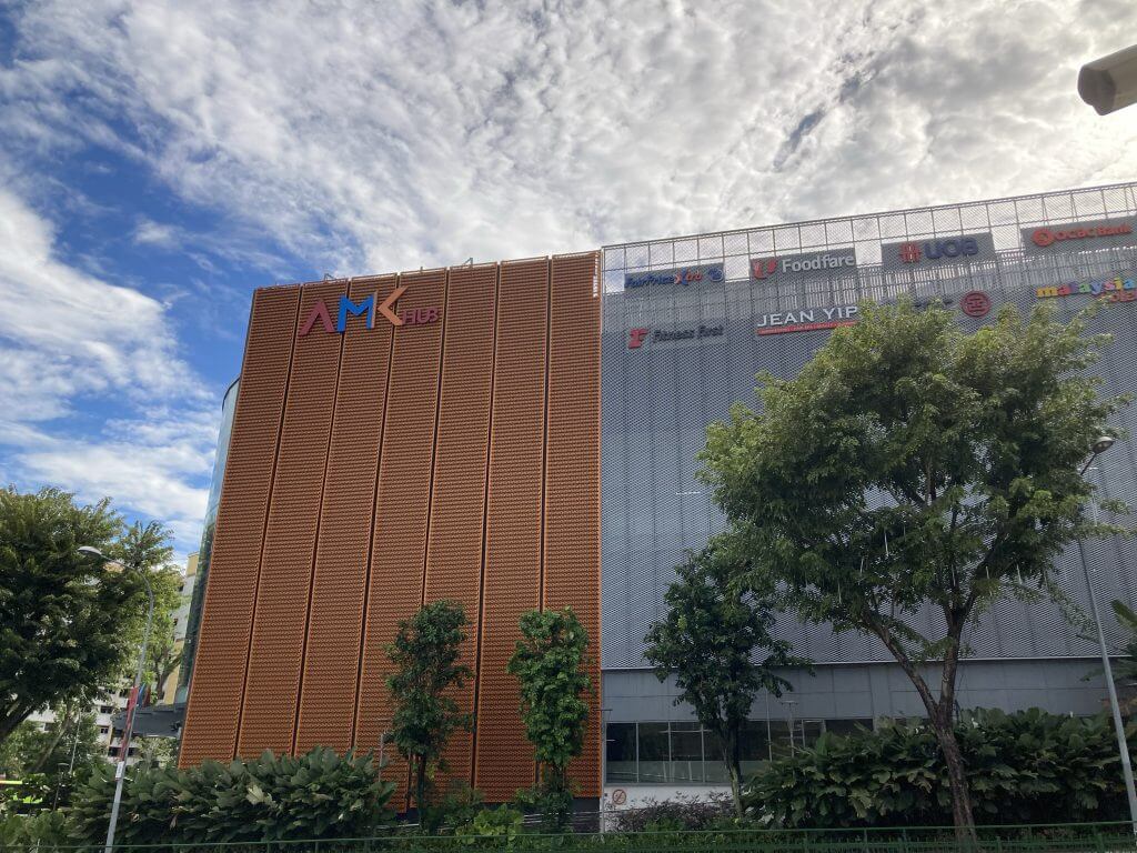
Health fanatics from the upcoming Belgravia Ace can take their choice from the multiple health and fitness services near house. There’s Health And Fitness First at AMK Center as well as Real Physical fitness, situated near Djitsun Shopping mall. They can likewise go to Yio Chu Kang Sports Centre by taking the train at the Yio Chu Kang MRT terminal.
Belgravia Ace at Belgravia Drive lies in D28, the central part of Singapore. This is created between Tong Eng Team This land will be redeveloped right into 107 Devices consists of 104 Semi-D + 3 Terraces. Purchasers of Belgravia Ace can anticipate elegant and also quality surfaces and also devices supplied by Tong Eng Group.
Belgravia Ace lies at Belgravia Drive, Ang Mo Kio precinct. It is in the Central Part of Singapore. Ang Mo Kio MRT Terminal is only mins’ drive away. Belgravia Ace Property landed is surrounded by numerous shopping malls as well as prominent institutions. As an example, Rosyth School is within 1km to Belgravia Ace. Belgravia Ace is just minutes drive to the close-by shopping malls such as Seletar Mall, Greenwich, AMK Center which can offer you a trendy and lively way of living. Besides retail shops, F&B, as well as amusement choices, there are also other services.
This future luxury landed is well attached to significant expressway like CTE as well as KPE as well as MCE that makes it extremely well connected to the other parts of Singapore. Enthusiastic proprietors of Belgravia Ace can choose to discover even more surrounding facilities with the Belgravia Ace location map.
This attractive landed building is surrounded by numerous fantastic facilities. There is a vast array of F&B and also buying centers are available in neighboring shopping centers like Greenwich V, Ferndale Mall, as well as The Seletar Shopping center. The growth is likewise within close closeness to the main road as well as significant expressway. Belgravia Ace is easy to gain access to via Central Expressway– CTE, Seletar Expressway– SLE, Tampines Expressway– TPE, as well as the future North-South Expressway (NSE ). For those homeowner that treasure the comfort of living in a landed residence with the included benefit of facilities, BELGRAVIA ACE is definitely made for you.
Belgravia Ace is a freehold strata landed houses constructing in Belgravia Drive Singapore. This property lies in Singapore’s Area 20, in the Seletar Hills Estate. Your homes are all oriented in a North South instructions, which will suit the majority of occupants. This house has an estate title. Fairview Developments Private Limited is the firm behind the task. They belong to the Tong Eng Event.
The total location of the land is predicted to be 136,562 square feet/12,678 square meters. The project’s forecasted TOP day is June 2023, with a delivery date of June 2026.
Anderson JC, Lycee Francais De Singapour (French International College), Rosyth School, Nanyang Polytechnic, as well as others are amongst the prestigious educational facilities in the area that will certainly be fairly practical for family members of school-aged youngsters.
In between the established Estate of Ang Mo Kio as well as the modern vibrant Seng Kang, residents at Belgravia Ace Singapore can experience world-class centers. This will enable homeowners to make use of the special eating as well as retail choices readily available. The Seletar Center, a brand-new shopping plaza with a number of dining establishments, fitness classes, restaurants, and facilities, is additionally close by. This will mean that citizens can obtain all of their food, purchasing, as well as various other needs from this place.
Belgravia Ace at Belgravia Drive lies in D28, the central part of Singapore. This is established between Tong Eng Group This land will be redeveloped into 107 Systems comprises 104 Semi-D + 3 Balconies. Buyers of Belgravia Ace can anticipate elegant and high quality surfaces and also appliances offered by Tong Eng Team.
Belgravia Ace is located at Belgravia Drive, Ang Mo Kio district. It is in the Central Part of Singapore. Ang Mo Kio MRT Station is just mins’ drive away. Belgravia Ace Freehold landed is bordered by numerous mall and prestigious colleges. As an example, Rosyth School is within 1km to Belgravia Ace.
Belgravia Ace is just mins drive to the neighboring shopping malls such as Seletar Shopping Mall, Greenwich, AMK Center which can offer you a stylish and also dynamic way of life. Besides retail shops, F&B, as well as home entertainment choices, there are likewise other features.
This approaching luxury landed is well connected to major expressway like CTE and also KPE and also MCE that makes it very well attached to the other parts of Singapore.
Hopeful proprietors of Belgravia Ace can select to discover more surrounding facilities with the Belgravia Ace place map.
Daily grocery stores shopping like NTUC, Sheng Siong and Cold Storage are close closeness to the Belgravia Ace that makes it really practical. Seletar Shopping Mall, The Greenwich Village, AMK Center offers fantastic dining as well as enjoyment choices. Homeowners can either drive or take public transport to Orchard Buying belt for much more options for eating as well as home entertainment.
The Midwood is a new growth situated at Hillview Riseby Hong Leong Holdings. which consist of 564 units in 2 29-storeys towers. The growth lies in the silent area of Hillview and also provide breathtaking views of the bordering plant. Iis situated just 300 meters from Hillview MRT Station. The Midwood Hong Leong was awarded under the Principle and Rate Profits tender which assess numerous tender proposals by developers and also is awarded not simply based on cost alone but various other elements such as boosting the social features of the immediate location. The Midwood is hotly opposed due to the absence of property space readily available for development. A special living experience awaits you at The Midwood at Hillview Surge.
The Midwood Condominium is also tactically situated with several shopping center situated around around. For example, the preferred hillV2 mall, The Rail Mall and Hillion Mall lies near to The Midwood Hillview Rise. The Midwood Hillview Apartment is likewise located close to Hillview as well as Bukit Batok Nature Get where plenty of exterior families are available for citizens to spend some top quality time with their household. The Midwood Hillview Rise will come with the existing Hillview MRT Terminal on the Midtown Line that takes you to the city straight. Additionally, for proprietors who are taking buses, there are numerous buses offered along Hillview Rise, Hillview Method as well as Dairy Farm Road. For owners that are taking a trip to the city, The Midwood Condominium is located right alongside Upper Bukit Timah Road and also. The Midwood Singapore is additionally near elite colleges such as CHIJ (Our Lady Queen of Peace), Saint Francis Methodist School and German European School Singapore (GESS).
The Midwood Apartment is being established by Intrepid Investments as well as Yard Estates, fully-owned systems of Hong Leong Holdings Limited as well as sister firm Hong Realty respectively. Hong Leong Holdings was developed in 1968 as the home financial investment and development arm of the Hong Leong Team. One of the earliest moving companies in the local realty scene, it has actually considering that expanded to become a major home player. Along the way it has earned a credibility as one of one of the most respected as well as relied on suppliers of homes in Singapore. Hong Leong Holdings handles 8 business jobs currently and also developed some 100 property projects covering the mid to premium array, including in several of the island’s most valued neighbourhoods. Going forward, it means to boost and expand its organization, while remaining to maintain the quality of work that it is understood for. Regarding the Hong Leong Group Singapore (” Hong Leong” or “the Team”).
The Hong Leong Group is among Singapore’s largest organization conglomerates. It is involved in a number of service areas in the Asia-Pacific, The United States And Canada and Europe. The Team’s 5 core business areas are home advancement and also investment, resort management as well as ownership, trade & sector, monetary solutions, as well as e-Business. On the residential or commercial property front, the Group is recognised as a key contributor to the neighborhood property scene. Via its 2 residential property arms, Hong Leong Holdings and also SGX-listed City Developments Limited (CDL), it holds the greatest residential land bank in Singapore beyond the federal government. So it is rarely shocking that the Team must rate as our largest developer of property jobs.
We have actually laid out some of its top information below: About 3 mins of walk to the Hillview MRT station In close proximity to appreciated colleges Situated right next to HillV2 Shopping mall A train quit away from Rail Shopping center A lot of nature parks as well as reserves around– including Bukit Timah as well as Bukit Batok A Hong Leong Limited residential or commercial property Full with a full-sized swimming pool, play area, and also cycling as well as strolling tracks.
Here’s another enhancement to Hong Leong Holdings long streak of properly designed and obtainable developments: The Midwood Condominium. As they have constantly done, the Hong Leong designers are understood for selling more than simply a roof over one’s head– they offer a way of life.
This time around, The Midwood Condominium, provides a quiet life set amongst the lush plants of Hillview Rise while still providing its homes fast access to the busy life in Singapore’s city. With it, you can conveniently escape from the hustle and bustle of the city as well as come home to one of Singapore’s desirable neighborhoods. The facilities of the condominium itself can be something to expect too. In fact, the authorities in the property market have actually taken notice of this new residential advancement, and they have actually provided Hong Leong acknowledgment under Concept and also Price revenue. Such distinction is offered to developers who exhibit a remarkably keen eye for enhancing the centers in its immediate location. When it comes to its dimension, the apartment itself covers the room of a tremendous 153,825 sq. ft. of land.
Resting regally in this apparently boundless area are 2 towering structures of each over 29 floorings. In due time, over 564 property owners and their families can call The Midwood new launch their home. Simply visualize: as a local of The Midwood Apartment, you’ll promptly be greeted with lavish greenery as well as a tranquil neighborhood as you step out of the building. If you find yourself in need of family products or simply wish to have a quiet supper with your close friends as well as loved ones, you can simply stroll to Hillv2 which is just a rock’s get rid of. You can quickly head to Marina Bay Sands as well as Chinatown by means of the Hillview MRT Terminal as well. All you have to do is take a simple 3-minute walk to the terminal from the condominium. The Midwood area as well as its facilities are genuinely second to none. If you want to learn more about this encouraging brand-new household growth, then go on reading.
The Midwood Condo Area map If there’s anything to be claimed about the Midwood developer, Hong Leong Holdings Limited, it’s that they are really one of the best in building way of lives. Check out: New Introduce Apartment In SG Near MRT (2020 ).
They don’t just develop cookie-cutter domestic buildings. Instead, they think about just what the marketplace requires and after that build that in a top-notch place. The Midwood Apartment 3D Map With Midwood Condo new launch, they have actually developed the excellent blend of a peaceful way of living in the metropolitan forest that is Singapore. They have actually taken care of to develop a residential property set against the background of the attractive vegetation of the city.
But while greenery might be synonymous to remote mountain-living, the Midwood Apartment is really fairly easily accessible in its own right. Simply 3 mins away is the Hillview MRT Terminal which can bring you to and from popular city sites such as Chinatown and Marina Bay Sands.
If you need to do stock up on materials or want to order a fast grub, The Rail Mall is simply a stop away also. The Midwood Condo really weds ease of access and living a quiet nature-filled life. It’s no wonder several wait for the Midwood Apartment launch day and the Midwood Condo Top date in 2023.
We have the designers Hong Leong Holdings (HLH) Restricted to say thanks to for this brand-new house. They are well-known as one of Singapore’s a lot of respected as well as desired developers. As one might expect from a firm with over 40 years of experience, they have actually currently accumulated a lot of finished jobs under their belts. As a matter of fact, they have actually completed over a hundred house buildings as well as 8 business developments. From the sheer quantity of their works alone, we know that these are the people that we can trust to develop our residences. Read about: Which should you pick, 99 Years Or Freehold– The Full Overview Also property owners outside of Singapore believe in the professionalism and trust of Hong Leong Holdings also. They have actually finished contracts with businesses from throughout the world– from Europe to Asia Pacific as well as in North America too. The profile of its developers alone has us excited for the Midwood.
Enduring in nature does not indicate that you will be losing out on the conveniences of the city– well, at the very least not with Midwood Condo. Here, you will certainly be much sufficient to appreciate a silent evening after work, but near adequate to be able to go out and also take pleasure in the mall or various other sources of entertainment also. Apart from the Hillv2 Shopping Centre, there’s the Rail Shopping center which is quickly one train stop away from the condo. Below, you’ll be able to appreciate a selection of cuisines and buy all the basic points you might require at home. If you’re trying to find an intriguing way to invest your weekend, you can most likely to Singapore Quarry simply next to it as well. This observatory permits you to completely welcome the natural beauty of the night skies. You might likewise such as: Ultimate Guide to Fixed Deposit If you prefer to invest even more time in nature, the Hillview location goes to the core of various nature parks draw as Bukit Batok and also Bukit Gombak as well. Getting around Singapore will certainly be simple for homeowners of the Midwood Apartment as well. There are several buses plying the location as well as the Hillview MRT Terminal is just a brief 3-minute walk from the apartment too. In terms of access as well as features, this residential or commercial property truly sets the ideal balance in between indulging in nature and delighting in one’s contemporary sensibilities. The Midwood Apartment to hillview MRT The Midwood Condominium Bukit Batok Town Park The Midwood Condominium near Bukit Timah.
Considering the Midwood Condo as a feasible financial investment chance? This prime piece of home can be your own for a reasonable cost too. If you’re planning on acquiring this residential property for rental fee, its ease of access alone can be your major selling factor. Review: Affordable Condominium for sale in Singapore In addition to, you’ll be considering high yields and strong returns if you manage to rent out your residential property to the ideal renters. The residential property itself will certainly have various formats to satisfy differing customer accounts– as well as you can discover more regarding this in the Midwood Condo layout or perhaps through the Midwood Condo showflat. Although the numbers need to be discussed by thinking about the Midwood cost amongst several things, the serenity as well as access of the residential property will simply generally be marketing the place for you. bukit batok plan of attack back Bukit Batok map Bukit Batok Bukit Batok.
The Midwood Apartment neighboring colleges If you’re intending on staying in Midwood Apartment with your kids, after that you won’t need to look much to obtain a top quality education and learning for them as well. The location itself is child-friendly with several play areas as well as day care facilities for the younger ones. For the older kids, you can have elite schools nearby such as Saint Francis Methodist Institution, CHIJ (Our Woman of Tranquility), as well as GESS (German European College Singapore). you might also like: Top 10 tips for getting involved in excellent institution in Singapore.
Midwood Apartment prides itself on its full features. From a full-size pool, spacious function rooms, as well as outside eating experiences, this property supplies features that are as considerable as they are convenient. There’s a checking out skies deck, an exterior exercise space, and also various other costs centers also. Read additionally: Property Taxes in Singapore Every one of the cycling paths and strolling trails in the area will certainly have you pedaling and also walking about in the direction of a healthy and balanced and also delighted lifestyle. The Midwood Condo house The Midwood Condo infinity.
The rates of the systems vary and will depend on the format as well as the dimension variety of your finding. A one-bedroom system will likely set you back at around $750,000 for the Midwood Apartment PSF. On the other hand, you can purchase a home of as much as 4 bedrooms at a cost for the Midwood PSF at $1.999 million. understand more regarding: TOP Projects in Singapore.
Certainly, the Midwood Condo is absolutely a financial investment in a way of life that lots of can only dream of. With its quiet area in an area with bountiful vegetation, you and your household will have the ability to totally leave the hustle and bustle of the city. And also when recharged, you can conveniently take a trip back to the city with simply an easy train or bus ride away also. Its critical area makes it feasible for you to balance restful nights while still enjoying the comforts of the city.
We hope that you’ve found out more via the Midwood testimonial. Ask us about this Hillview property and also we’ll be greater than delighted to walk answer any inquiries or provide you with The Midwood Condo Pamphlet.